Citation:
Abstract:
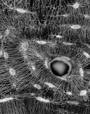
Fish represent the most diverse and numerous of the vertebrate clades. In contrast to the bones of all tetrapods and evolutionarily primitive fish, many of the evolutionarily more advanced fish have bones that do not contain osteocytes. Here we use a variety of imaging techniques to show that anosteocytic fish bone is composed of a sequence of planar layers containing mainly aligned collagen fibrils, in which the prevailing principal orientation progressively spirals. When the sequence of fibril orientations completes a rotation of around 180, a thin layer of poorly oriented fibrils is present between it and the next layer. The thick layer of aligned fibrils and the thin layer of non-aligned fibrils constitute a lamella. Although both basic components of mammalian lamellar bone are found here as well, the arrangement is unique, and we therefore call this structure lamellated bone. We further show that the lamellae of anosteocytic fish bone contain an array of dense, small-diameter (1-4 mu m) bundles of hypomineralized collagen fibrils that are oriented mostly orthogonal to the lamellar plane. Results of mechanical tests conducted on beams from anosteocytic fish bone and human cortical bone show that the fish bones are less stiff but much tougher than the human bones. We propose that the unique lamellar structure and the orthogonal hypomineralized collagen bundles are responsible for the unusual mechanical properties and mineral distribution in anosteocytic fish bone. (C) 2014 Acta Materialia Inc. Published by Elsevier Ltd. All rights reserved.

