Yair R, Shahar R, Uni Z.
In ovo feeding with minerals and vitamin D-3 improves bone properties in hatchlings and mature broilers. POULTRY SCIENCE [Internet]. 2015;94 (11) :2695-2707.
Publisher's VersionAbstractThe objective of this study was to examine the effect of in ovo feeding (IOF) with inorganic minerals or organic minerals and vitamin D-3 on bone properties and mineral consumption. Eggs were incubated and divided into 4 groups: IOF with organic minerals, phosphate, and vitamin D-3 (IOF-OMD); IOF with inorganic minerals and phosphate (IOF-IM); sham; and non-treated controls (NTC). IOF was performed on embryonic day (E) 17; tibiae and yolk samples were taken on E19 and E21. Post-hatch, only chicks from the IOF-OMD, sham, and NTC were raised, and tibiae were taken on d 10 and 38. Yolk mineral content was examined by inductively coupled plasma spectroscopy. Tibiae were tested for their whole-bone mechanical properties, and mid-diaphysis bone sections were indented in a micro-indenter to determine bone material stiffness (Young's modulus). Micro-computed tomography (mu CT) was used to examine cortical and trabecular bone structure. Ash content analysis was used to examine bone mineralization. A latency-to-lie (LTL) test was used to measure standing ability of the d 38 broilers. The results showed that embryos from both IOF-OMD and IOF-IM treatments had elevated Cu, Mn, and Zn amounts in the yolk on E19 and E21 and consumed more of these minerals (between E19 and E21) in comparison to the sham and NTC. On E21, these hatchlings had higher whole-bone stiffness in comparison to the NTC. On d 38, the IOF-OMD had higher ash content, elevated whole-bone stiffness, and elevated Young's modulus (in males) in comparison to the sham and NTC; however, no differences in standing ability were found. Very few structural differences were seen during the whole experiment. This study demonstrates that mineral supplementation by in ovo feeding is sufficient to induce higher mineral consumption from the yolk, regardless of its chemical form or the presence of vitamin D-3. Additionally, IOF with organic minerals and vitamin D-3 can increase bone ash content, as well as stiffness of the whole bone and bone material in the mature broiler, but does not lead to longer LTL.
 in_ovo_feeding_with_minerals_and_vitamin_d-3_improves_bone_properties_in_hatchlings_and_mature_broilers.pdf
 Stern T, Aviram R, Rot C, Galili T, Sharir A, Achrai NK, Keller Y, Shahar R, Zelzer E.
Isometric Scaling in Developing Long Bones Is Achieved by an Optimal Epiphyseal Growth Balance. PLOS BIOLOGY [Internet]. 2015;13 (8).
Publisher's VersionAbstractOne of the major challenges that developing organs face is scaling, that is, the adjustment of physical proportions during the massive increase in size. Although organ scaling is fundamental for development and function, little is known about the mechanisms that regulate it. Bone superstructures are projections that typically serve for tendon and ligament insertion or articulation and, therefore, their position along the bone is crucial for musculoskeletal functionality. As bones are rigid structures that elongate only from their ends, it is unclear how superstructure positions are regulated during growth to end up in the right locations. Here, we document the process of longitudinal scaling in developing mouse long bones and uncover the mechanism that regulates it. To that end, we performed a computational analysis of hundreds of three-dimensional micro-CT images, using a newly developed method for recovering the morphogenetic sequence of developing bones. Strikingly, analysis revealed that the relative position of all superstructures along the bone is highly preserved during more than a 5-fold increase in length, indicating isometric scaling. It has been suggested that during development, bone superstructures are continuously reconstructed and relocated along the shaft, a process known as drift. Surprisingly, our results showed that most superstructures did not drift at all. Instead, we identified a novel mechanism for bone scaling, whereby each bone exhibits a specific and unique balance between proximal and distal growth rates, which accurately maintains the relative position of its superstructures. Moreover, we show mathematically that this mechanism minimizes the cumulative drift of all superstructures, thereby optimizing the scaling process. Our study reveals a general mechanism for the scaling of developing bones. More broadly, these findings suggest an evolutionary mechanism that facilitates variability in bone morphology by controlling the activity of individual epiphyseal plates.
 isometric_scaling_in_developing_long_bones_is_achieved_by_an_optimal_epiphyseal_growth_balance.pdf
 Jimenez-Palomar I, Shipov A, Shahar R, Barber AH.
Mechanical behavior of osteoporotic bone at sub-lamellar length scales. FRONTIERS IN MATERIALS. 2015;13 :1-7.
AbstractOsteoporosis is a disease known to promote bone fragility but the effect on the mechanical
properties of bone material, which is independent of geometric effects, is particularly
unclear. To address this problem, micro-beams of osteoporotic bone were prepared using
focused ion beam microscopy and mechanically tested in compression using an atomic
force microscope while observing them using in situ electron microscopy.This experimental
approach was shown to be effective for measuring the subtle changes in the mechanical
properties of bone material required to evaluate the effects of osteoporosis. Osteoporotic
bone material was found to have lower elastic modulus and increased strain to failure
when compared to healthy bone material, while the strength of osteoporotic and healthy
bone was similar. Surprisingly, the increased strain to failure for osteoporotic bone material
provided enhanced toughness relative to the control samples, suggesting that lowering of
bone fragility due to osteoporosis is not defined by material performance. A mechanism is
suggested based on these results and previous literature that indicates degradation of the
organic material in osteoporosis bone is responsible for resultant mechanical properties.
 mechanical_behavior_of_osteoporotic_bone_at_sub-lamellar_length_scales.pdf
 Jimenez-Palomar I, Shipov A, Shahar R, Barber AH.
Structural orientation dependent sub-lamellar bone mechanics. JOURNAL OF THE MECHANICAL BEHAVIOR OF BIOMEDICAL MATERIALS [Internet]. 2015;52 :63-71.
Publisher's VersionAbstractThe lamellar unit is a critical component in defining the overall mechanical properties of bone. In this paper, micro-beams of bone with dimensions comparable to the lamellar unit were fabricated using focused ion beam (FIB) microscopy and mechanically tested in bending to failure using atomic force microscopy (AFM). A variation in the mechanical properties, including elastic modulus, strength and work to fracture of the micro-beams was observed and related to the collagen fibril orientation inferred from back-scattered scanning electron microscopy (SEM) imaging. Established mechanical models were further applied to describe the relationship between collagen fibril orientation and mechanical behaviour of the lamellar unit. Our results highlight the ability to measure mechanical properties of discrete bone volumes directly and correlate with structural orientation of collagen fibrils. (C) 2015 Elsevier Ltd. All rights reserved.
 structural_orientation_dependent_sub-lamellar_bone_mechanics.pdf
 Atkins A, Milgram J, Weiner S, Shahar R.
The response of anosteocytic bone to controlled loading. JOURNAL OF EXPERIMENTAL BIOLOGY [Internet]. 2015;218 (22) :3559-3569.
Publisher's VersionAbstractThe bones of the skeleton of most advanced teleost fish do not contain osteocytes. Considering the pivotal role assigned to osteocytes in the process of modeling and remodeling (the adaptation of external and internal bone structure and morphology to external loads and the repair of areas with micro-damage accumulation, respectively) it is unclear how, and even whether, their skeleton can undergo modeling and remodeling. Here, we report on the results of a study of controlled loading of the anosteocytic opercula of tilapia (Oreochromis aureus). Using a variety of microscopy techniques we show that the bone of the anosteocytic tilapia actively adapts to applied loads, despite the complete absence of osteocytes. We show that in the directly loaded area, the response involves a combination of bone resorption and bone deposition; we interpret these results and the structure of the resultant bone tissue to mean that both modeling and remodeling are taking place in response to load. We further show that adjacent to the loaded area, new bone is deposited in an organized, layered manner, typical of a modeling process. The material stiffness of the newly deposited bone is higher than that of the bone which was present prior to loading. The absence of osteocytes requires another candidate cell for mechanosensing and coordinating the modeling process, with osteoblasts seeming the most likely candidates.
 the_response_of_anosteocytic_bone_to_controlled_loading.pdf
 Atkins A, Reznikov N, Ofer L, Masic A, Weiner S, Shahar R.
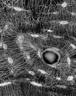
The three-dimensional structure of anosteocytic lamellated bone of fish. ACTA BIOMATERIALIA [Internet]. 2015;13 :311-323.
Publisher's VersionAbstractFish represent the most diverse and numerous of the vertebrate clades. In contrast to the bones of all tetrapods and evolutionarily primitive fish, many of the evolutionarily more advanced fish have bones that do not contain osteocytes. Here we use a variety of imaging techniques to show that anosteocytic fish bone is composed of a sequence of planar layers containing mainly aligned collagen fibrils, in which the prevailing principal orientation progressively spirals. When the sequence of fibril orientations completes a rotation of around 180, a thin layer of poorly oriented fibrils is present between it and the next layer. The thick layer of aligned fibrils and the thin layer of non-aligned fibrils constitute a lamella. Although both basic components of mammalian lamellar bone are found here as well, the arrangement is unique, and we therefore call this structure lamellated bone. We further show that the lamellae of anosteocytic fish bone contain an array of dense, small-diameter (1-4 mu m) bundles of hypomineralized collagen fibrils that are oriented mostly orthogonal to the lamellar plane. Results of mechanical tests conducted on beams from anosteocytic fish bone and human cortical bone show that the fish bones are less stiff but much tougher than the human bones. We propose that the unique lamellar structure and the orthogonal hypomineralized collagen bundles are responsible for the unusual mechanical properties and mineral distribution in anosteocytic fish bone. (C) 2014 Acta Materialia Inc. Published by Elsevier Ltd. All rights reserved.
 the_three-dimensional_structure_of_anosteocytic_lamellated_bone_of_fish.pdf
