Publications

2021
Polycations and polyanions in SARS-CoV-2 infection
Ginsburg I, Fibach E. Polycations and polyanions in SARS-CoV-2 infection. Medical Hypotheses Volume 146 [Internet]. 2021;146. Publisher's VersionAbstract

We hypothesize that polycations, such as nuclear histones, released by neutrophils COVID-19 aggravate COVID-19 by multiple mechanisms: (A) Neutralization of the electrostatic repulsion between the virus particles and the cell membrane, thereby enhancing receptor-mediated entry. (B) Binding to the virus particles, thereby inducing opsonin-mediated endocytosis. (C) Adding to the cytotoxicity, in conjunction with oxidants, cytokines and other pro-inflammatory substances secreted by cells of the innate immunity system. These effects may be alleviated by the administration of negatively charged polyanions such as heparins and heparinoids.

1-s2.0-s0306987720333612-main.pdf
2020
Longitudinal patterns of cytokine expression at the individual level in humans after laparoscopic sleeve gastrectomy
Trahtemberg U, Darawshe F, Elazary R, Ginsburg I, Beil M, van Heerden PV, Sviri S. Longitudinal patterns of cytokine expression at the individual level in humans after laparoscopic sleeve gastrectomy. Journal of Cellular and Molecular Medicine [Internet]. 2020;24 (12) :6622-6633. Publisher's VersionAbstract

The study of the human response to injury has been hampered by the inherent heterogeneity in the models and methods used. By studying a standard injury longitudinally, using individual patient‐level analysis, we endeavoured to better describe its dynamics. We analysed clinical variables, clinical laboratory and plasma cytokines from 20 patients at five time points. Clustering analysis showed two prototype patterns of cytokine behaviour: a concordant type, where cytokines behave the same way for all patients (notably IL‐0 and TNFα), and a variable type, where different patterns of expression are seen for different patients (notably IL‐8, IL‐6 and IL‐1RA). Analysis of the cytokines at the individual patient‐level showed a strong four‐way correlation between IL‐1RA, GCSF, MIP‐1β and MCP‐1. As it holds for most patients and not just on average, this suggests that they form a network which may play a central role in the response to gastro‐intestinal injuries in humans. In conclusion, the longitudinal analysis of cytokines in a standard model allowed the identification of their underlying patterns of expression. We propose that the two prototype patterns shown may reflect the mechanism that separates the common and individual aspects of the injury response.

A Novel Concept May Explain How Immune Complexes Interact with Highly Cationic Histones Released by Activated Neutrophils Nets Act in Synergy with the Plethora of Neutrophils Pro-Inflammatory Agonists Leading to the Development of Autoimmune Nephritis -A
Ginsburg I, Koren E, Dov IB. A Novel Concept May Explain How Immune Complexes Interact with Highly Cationic Histones Released by Activated Neutrophils Nets Act in Synergy with the Plethora of Neutrophils Pro-Inflammatory Agonists Leading to the Development of Autoimmune Nephritis -A . Journal of Clinical Nephrology & Kidney Diseases [Internet]. 2020;5 (1). Publisher's VersionAbstract


Recent studies have pointed out that highly cationic histones released by PMNs netosis may be major agents in autoimmune lupus since they have high affinity to various kidney sites and can be expected to play a key role in autoimmune glomerular disease.

Similarly to antibodies, cationic peptides such a nuclear histone can also act as potent opsonic agents capable of binding by strong electrostatic forces to negatively charged domains in immune complexes and in complement components resulting in their endocytosis and deposition in various parts of the kidney. It is also proposed that to prevent such events, highly anionic heparin and heparinoids, may be effective drugs since these may effectively neutralize histones activities but provided that agents such as steroids, methotrexate and colchicine, all potent inhibitors of neutrophils functions, and antibodies to TH1 cytokines be essential to treat nephritis and to prevent kidney failure. However, the main cause of kidney damage is eventually caused by the plethora of toxic pro inflammatory agents delivered by activated neutrophils and macrophages.


nephritis.pdf
In Lupus Erythematosus the Deposition of Immune Complexes in Tissues Is Mediated via Nuclear Histones Released by Neutrophils Nets but the Main Damage to Hosts Tissues Is Caused by the Plethora of Toxic Pro Inflammatory Agents Released by Activated Neutro
Ginsburg I. In Lupus Erythematosus the Deposition of Immune Complexes in Tissues Is Mediated via Nuclear Histones Released by Neutrophils Nets but the Main Damage to Hosts Tissues Is Caused by the Plethora of Toxic Pro Inflammatory Agents Released by Activated Neutro. Open Journal of Rheumatology and Autoimmune Diseases [Internet]. 2020;10 (2). Publisher's VersionAbstract

The present study offers a novel approach that may explain the mechanisms of pathogenicity of the auto immune destructive disorder, Lupus Erythematosus. It is proposed that deposition of immune complexes and complement components in tissue is mediated by highly cationic histones released from neutrophils nets the phenomenon of netosis. Histones act a potent opsonic factor similar to antibodies which interact by strong electrostatic forces with negatively-charged domains in immune complexes and complements facilitating their deposition and also their internalization by hosts’ cells. However, the main cause of cell and tissue damage in Lupus is inflicted by the plethora of toxic pro inflammatory agonists released by neutrophils and by macrophages recruited to inflamed tissues by cytokines. The melioration of tissue damage may be initiated by highly anionic heparins, which neutralizes histones’ action if also combined with steroids, colchicin and methtorxate as well as by other agents which retard leukocytes migration and functions.

ojra_2020052815445046.pdf
A novel aspect may explain the mechanisms of pathogenicity of rheumatic fever, a multifactorial, autoimmune, infectious and inflammatory disorder which “licks the joints and bites the heart”: A working hypothesis
Ginsburg I, Feldman M. A novel aspect may explain the mechanisms of pathogenicity of rheumatic fever, a multifactorial, autoimmune, infectious and inflammatory disorder which “licks the joints and bites the heart”: A working hypothesis. Medical Hypotheses [Internet]. 2020;144. Publisher's VersionAbstract

A novel hypothesis is presented to explain the pathogenesis of the multifactorial autoimmune disorder rheumatic fever (RF). It involves a synergistic interaction among streptococcal toxins, their cell wall components, M protein, immune complexes, complement components, cationic histones. These agents can act with cationic histones released by neutrophils during NETosis and bacteriolysis and can function as opsonic agents possessing properties similar to antibodies. Cationic histones can interact by strong electrostatic forces with negatively- charged domains on immune complexes and complement components. This allows their deposition and endocytosis in the myocardium, the heart valves, and in the joints. However, the main cause of cell and tissue damage observed in RF is due to a synergism among the plethora of pro-inflammatory substances released by activated neutrophils and macrophages. Cell damage may be mitigated to some extent by anionic heparins, heparinoids, and by anti-inflammatory drugs such as corticosteroids which counteract neutrophils and macrophage chemotaxis induced by cytokines.

1-s2.0-s030698772031402x-main.pdf
2019
A Novel Approach That May Explain the Role of Staphylococcus aureus, Polycations, Neutrophils Pro-Inflammatory Agonists and the Bacteriolysis and Auto Immune Phenomena as Possible Major Events in the Pathogenesis of Atopic Dermatitis: A Working Hypothesis
Ginsburg I, Koren E. A Novel Approach That May Explain the Role of Staphylococcus aureus, Polycations, Neutrophils Pro-Inflammatory Agonists and the Bacteriolysis and Auto Immune Phenomena as Possible Major Events in the Pathogenesis of Atopic Dermatitis: A Working Hypothesis. Journal of Clinical & Experimental Dermatology Research [Internet]. 2019;10 (5). Publisher's VersionAbstract


The aim of the present short communication is to shed a novel light on the auto immune disorder atopic dermatitis by discussing the possible role played by the plethora of toxic agents released by  Staphylococcus aureus which can act in a tight synergism with neutrophils derived cationic polyelectrolytes  as related to the pathogenesis of atopic dermatitis (AD) [1,2]. This disorder results in  inflammation of the  skin characterized by itchiness,  red  skin,  a  rash,  by   the  accumulations  of  large numbers of Staphylococcus  aureus  their toxins [2] and pro- inflammatory agents secreted by migrating neutrophiles [3], are considered the main cause of AD pathogenicity.


2155-9554-10-508_copy.pdf
Is Tissue Damage caused in Group A Streptococcal Infections Augmented by Synergizing with Neutrophils’ Pro-inflammatory Products?
Ginsburg I, Koren E. Is Tissue Damage caused in Group A Streptococcal Infections Augmented by Synergizing with Neutrophils’ Pro-inflammatory Products?. Microbiology & Infectious Diseases [Internet]. 2019;2019; 3 (1) :1-6. Publisher's VersionAbstract

Catalase-negative penicillin-sensitive group A hemolytic streptococci (GAS) are multifactorial microorganisms, which do not produce a unique damage-associated molecular patterns which if effectively neutralized might effectively stop their pathogenicity. GAS is involved in the pathogenicity of pharangitis, tonsillitis, rheumatic fever, arthritis, necrotizing fasciitis (NF), toxic shock syndrome and also in sepsis. GAS-induced NF is quite a rare but dangerous and deadly infection, which most commonly occurs in the arms, legs and abdominal wall and is fatal in 30%-40% of cases. GAS, which possess surface capsular polysaccharide and antigenic M and T proteins, arrive at the inflammatory areas by generating spreading factors such as hyaluronidase, DNase and streptokinaseactivated plasmin. GAS can spread in tissues and avidly adhere to membranes of target cells to deliver a nonimmunogenic cell bound hemolysin (CBH) upon cells’ membrane phospholipids to induce a penetrating membrane damage (“a kiss of Death”). Two additional potent extracellular hemolysins, Streptolysin O (SLO) and a nonimmunogenic streptolysin S (SLS) produced can injure neutrophils (PMNs), which are recruited to the infected sites in large numbers. However, PMNs can engage in phagocytosis and also undergo activation to release various proinflammatory agents including NADPH-generated superoxide which dismutates to H2 O2 and with myeloperoxidase (MPO) which forms toxic HOCl upon interaction with halides. Activated PMNs also deliver highly cationic peptides such as LL37, cationic elastase, cathepsins and nuclear histone, which interact electrostatically with negatively-charged membrane sites forming membrane lesions. PMNs also secrete many acid hydrolases, several Th1 cytokines and chemokines, which recruit more PMNs. Similarly, to beta-lactams antibiotics, cationic peptides can also activate bacteriolysis and trigger the release of the pro-inflammatory agents lipoteichoic acid (LTA) and peptidoglycan (PPG). We hereby propose that in infectious and inflammatory sites GAS and PMNs exo-products and also microbial cellwall structures might all act synergistically to cause cell and tissue damage. Cell damage might be ameliorated by appropriate cocktails of anti-inflammatory agents. also, containing highly negatively charged heparin 23.

streop_papaer_.pdf
Pro-inflammatory agents released by pathogens, dying host cells, and neutrophils act synergistically to destroy host tissues: a working hypothesis
Ginsburg I, Koren M, Varani J. Pro-inflammatory agents released by pathogens, dying host cells, and neutrophils act synergistically to destroy host tissues: a working hypothesis. Journal of Inflammation Research [Internet]. 2019;12 :35-47. Publisher's VersionAbstract

Abstract: We postulate that the extensive cell and tissue damage inflicted by many infectious, inflammatory and post-inflammatory episodes is an enled result of a synergism among the invading microbial agents, host neutrophils and dead and dying cells in the nidus. Microbial toxins and other metabolites along with the plethora of pro-inflammatory agents released from activated neutrophils massively recruited to the infectious sites and high levels of cationic histones, other cationic peptides, proteinases and Th1 cytokines released from activated polymorphonuclear neutrophils (PMNs) and from necrotized tissues may act in concert (synergism) to bring about cell killing and tissue destruction. Multiple, diverse interactions among the many potential pro-inflammatory moieties have been described in these complex lesions. Such infections are often seen in the skin and aerodigestive tract where the tissue is exposed to the environment, but can occur in any tissue. Commonly, the tissue-destructive infections are caused by group A streptococci, pneumococci, Staphylococcus aureus, meningococci, Escherichia coli and Shigella, although many other microbial species are seen on occasion. All these microbial agents are characterized by their ability to recruit large numbers of PMNs. Given the complex nature of the disease process, it is proposed that, to treat these multifactorial disorders, a “cocktail” of anti-inflammatory agents combined with non-bacteriolytic antibiotics and measures to counteract the critical toxic role of cationic moieties might prove more effective than a strategy based on attacking the bacteria alone.

jir-190007-pro-inflammatory-agents-released-by-pathogens-by-dying-host-012219.pdf
Polyphenols Inhibit Candida albicans and Streptococcus mutans Biofilm Formation.
Farkash Y, Feldman M, Ginsburg I, Steinberg D, Shalish M. Polyphenols Inhibit Candida albicans and Streptococcus mutans Biofilm Formation. Dentistry Journal [Internet]. 2019;7 (2). Publisher's VersionAbstract

Background:Streptococcus mutans (S. mutans) and Candida albicans (C. albicans) are two major contributors to dental caries. They have a symbiotic relationship, allowing them to create an enhanced biofilm. Our goal was to examine whether two natural polyphenols (Padma hepaten (PH) and a polyphenol extraction from green tea (PPFGT)) could inhibit the caries-inducing properties of S. mutans and C. albicans. Methods: Co-species biofilms of S. mutans and C. albicans were grown in the presence of PH and PPFGT. Biofilm formation was tested spectrophotometrically. Exopolysaccharides (EPS) secretion was quantified using confocal scanning laser microscopy. Biofilm development was also tested on orthodontic surfaces (Essix) to assess biofilm inhibition ability on such an orthodontic appliance. Results: PPFGT and PH dose-dependently inhibited biofilm formation without affecting the planktonic growth. We found a significant reduction in biofilm total biomass using 0.625 mg/mL PPFGT and 0.16 mg/mL PH. A concentration of 0.31 mg/mL PPFGT and 0.16 mg/mL PH inhibited the total cell growth by 54% and EPS secretion by 81%. A reduction in biofilm formation and EPS secretion was also observed on orthodontic PVC surfaces. Conclusions: The polyphenolic extractions PPFGT and PH have an inhibitory effect on S. mutans and C. albicans biofilm formation and EPS secretion.

dentistry-07-00042-v2.pdf
2018
Effect of fixed orthodontic appliances on nonmicrobial salivary parameters.
Zogakis IP, Koren E, Gorelik S, Ginsburg I, Shalish M. Effect of fixed orthodontic appliances on nonmicrobial salivary parameters. The Angle Orthodontist [Internet]. 2018;88 (6) :806-811. Publisher's VersionAbstract

Objectives: To examine possible changes in the levels of salivary antioxidants, C-reactive protein (CRP), cortisol, pH, proteins, and blood in patients treated with fixed orthodontic appliances. Materials and Methods: Salivary samples from 21 orthodontic patients who met specific inclusion criteria were collected before the beginning of orthodontic treatment (T0; baseline), 1 hour after bonding (T1), and 4–6 weeks after bonding (T2). Oxidant-scavenging ability (OSA) was quantified using a luminol-dependent chemiluminescence assay. Cortisol and CRP levels were measured using immunoassay kits. pH levels and presence of proteins and blood in the samples were quantified using strip-based tests. Results: A significant decrease in salivary pH was observed after bonding (P ¼ .013). An increase in oxidant-scavenging abilities during orthodontic treatment was detected, but the change was not statistically significant. Cortisol and CRP levels slightly increased after bonding, but the difference was small without statistical significance. Changes in the presence of proteins and blood were also insignificant. Conclusions: Exposure to fixed orthodontic appliances did not show a significant effect on salivary parameters related to inflammation or stress, with the exception of a significant but transient pH decrease after bonding. (Angle Orthod. 2018;88:806–811.)

111317-773.1.pdf
Green Tea Polyphenols and Padma Hepaten Inhibit Candida albicansBiofilm Formation
Farkash Y, Feldman M, Ginsburg I, Steinberg D, Shalish M. Green Tea Polyphenols and Padma Hepaten Inhibit Candida albicansBiofilm Formation. Evidence-Based Complementary and Alternative Medicine. 2018;2018.Abstract

Candida albicans (C. albicans) is the most prevalent opportunistic human pathogenic fungus and can cause mucosal membrane infections and invade the blood. In the oral cavity, it can ferment dietary sugars, produce organic acids and therefore has a role in caries development. In this study, we examined whether the polyphenol rich extractions Polyphenon from green tea (PPFGT) and Padma Hepaten (PH) can inhibit the caries-inducing properties of C. albicans. Biofilms of C. albicans were grown in the presence of PPFGT and PH. Formation of biofilms was tested spectrophotometrically after crystal violet staining. Exopolysaccharides (EPS) secretion was quantified using confocal scanning laser microscopy (CSLM). Treated C. albicans morphology was demonstrated using scanning electron microscopy (SEM). Expression of virulence-related genes was tested using qRT-PCR. Development of biofilm was also tested on an orthodontic surface (Essix) to assess biofilm inhibition ability on such appliances. Both PPFGT and PH dose-dependently inhibited biofilm formation, with no inhibition on planktonic growth. The strongest inhibition was obtained using the combination of the substances. Crystal violet staining showed a significant reduction of 45% in biofilm formation using a concentration of 2.5mg/ml PPFGT and 0.16mg/ml PH. A concentration of 1.25 mg/ml PPFGT and 0.16 mg/ml PH inhibited candidal growth by 88% and EPS secretion by 74% according to CSLM. A reduction in biofilm formation and in the transition from yeast to hyphal morphotype was observed using SEM. A strong reduction was found in the expression of hwp1, eap1, and als3 virulence associated genes. These results demonstrate the inhibitory effect of natural PPFGT polyphenolic extraction on C. albicans biofilm formation and EPS secretion, alone and together with PH. In an era of increased drug resistance, the use of phytomedicine to constrain biofilm development, without killing host cells, may pave the way to a novel therapeutic concept, especially in children as orthodontic patients.

ecam2018-1690747.pdf
The Pathogenesis of Sepsis: “If We Cannot beat them Alone Join Them?”
Korem M, Koren E, Ginsburg I. The Pathogenesis of Sepsis: “If We Cannot beat them Alone Join Them?”. International Journal of Microbiology & Infectious Diseases [Internet]. 2018;2 (3) :1-5. Publisher's VersionAbstract


 Sepsis and septic shock are probably the least understood human disorders which worldwide take the lives of millions of patients. Sepsis may be defined as a multifactorial synergistic phenomenon where no unique damage-associated molecular patterns –alarming is identified which if successfully neutralized, might mitigate and protects against death in sepsis. 

Microorganisms which invade the blood stream may activate neutrophils to adhere to endothelial cells and to form oxidant – dependent nets rich in highly toxic nuclear histones claimed to be the main cause of death in sepsis due to the dysregulation of endothelial functions. However, the histone saga was recently critically debated since high levels circulating histones are also found in many clinical disorders unrelated to sepsis, therefore, histones may not be considered as a unique damage-associated molecular patterns- alarming but as additional markers of severe cell damage. 

We hereby argue that the main cause of tissue damage in sepsis may be an end result of a synergism between the numerous neutrophils pro inflammatory agents and the multiplicity of similar pro inflammatory agents generated by hemolytic steptoccocci and by additional pathogenic microorganism which recruit large numbers PMNs to the inflammatory sites. It is recommended that in sepsis caused by hemolytic streptococci and by additional toxigenic bacteria, a use of cocktails of antagonists might be more beneficial therapeutic strategies and this in view of the total failure to treat sepsis only by administrations of single antagonists. Also, targeting PMNs by immunological strategies should be sought for, to mitigate synergies between leukocytes and microbial cells.


the-pathogenesis-of-sepsis-if-we-cannot-beat-them-alone-join-them-388.pdf
Bacteriolysis – a mere laboratory curiosity?
Ginsburg I, Koren E. Bacteriolysis – a mere laboratory curiosity?. Critical Reviews in Microbiology [Internet]. 2018. Publisher's VersionAbstract


The role of bacteriolysis in the pathophysiology of microbial infections dates back to 1893 when

Buchner and Pfeiffer reported for the first time the lysis of bacteria by immune serum and related

this phenomenon to the immune response. Later on, basic anti-microbial peptides and certain

beta-lactam antibiotics have been shown not only to kill microorganisms but also to induce bacteriolysis

and the release of cell-wall components.

In 2009, a novel paradigm was offered suggesting that the main cause of death in sepsis is due

to the exclusive release from activated human phagocytic neutrophils (PMNs) traps adhering

upon endothelial cells of highly toxic nuclear histone. Since activated PMNs also release a plethora

of pro-inflammatory agonists, it stands to reason that these may act in synergy with histone

to damage cells. Since certain beta lactam antibiotics may induce bacteriolysis, it is questioned

whether these may aggravate sepsis patient's condition. Enigmatically, since the term bacteriolysis

and its possible involvement in sepsis is hardly ever mentioned in the extensive clinical

articles and reviews dealing with critical care, we hereby aim to refresh the concept of bacteriolysis

and its possible role in the pathogenesis of post infectious sequelae.


5_22_2018_bacterioly.pdf
2017
Are histones real pathogenic agents in sepsis?
Ginsburg I, Koren E. Are histones real pathogenic agents in sepsis?. NATURE REVIEWS - IMMUNOLOGY [Internet]. 2017. Publisher's VersionAbstract


We read with interest the recent Review article by van der Poll et al.1(The immunopathology of sepsis and potential therapeutic targets. Nat. Rev. Immunol. 17, 407–420 (2017)).This Review article describes various immunopathological aspects of sepsis and relevant targets as potential therapeutics. Unfortunately, we feel the authors failed to acknowledge highly relevant published data related to the possible pathogenic role of histones in sepsis.

In 2009, a paper by Xu et al.2, published in Nature Medicine, claimed that the main cause of death in sepsis is the release of highly toxic histones from neutrophils, possibly from those activated to make neutrophil extracellular traps3. Xu and co-workers also showed that the toxicity of histones could be abolished by either heparin, activated protein C or antibodies to histones. However, despite being an important new insight, this study was not cited in the Review by van der Poll and colleagues. Since this study, several other papers have been published showing high levels of circulating histones in many clinical disorders unrelated to sepsis3,4,5,6,7. This has led to the suggestion that histones are not unique inflammation-inducing alarmins (also known as damage-associated molecular patterns (DAMPs)) but are actually markers of cell damage8,9. Notably, in the context of sepsis, highly toxic cationic histones may function not alone but in synergy with oxidants and a range of pro-inflammatory agonists that are also released from activated neutrophils10,11,12,13. Again, none of these publications was acknowledged in the Review by van der Poll and colleagues.

We believe this important information on the possible role of histones in sepsis should have been acknowledged in this Review to encourage unbiased reporting and scholarly debate14.


are_histones_real_pathogenic_agents_koren_nri_.2017.156_author_proof.pdf
Tissue damage in post infectious sequelae is caused by a synergism between microbial and neutrophils-derived agonists: a concern for a disregard for already published data
Ginsburg I, Koren E, Van Heerden V. Tissue damage in post infectious sequelae is caused by a synergism between microbial and neutrophils-derived agonists: a concern for a disregard for already published data. Journal of Emerging and Rare Diseases [Internet]. 2017;1 (1) :1-3. Publisher's VersionAbstract

Post infectious sequelae such as sepsis and septic shock are poorly understood and annually take the lives of millions over the world. Severe microbial infections caused by Gram Positive and Gram Negative bacteria and by fungi are the main causes, which are aggravated by the rapid development of antibiotic resistance. It is unfortunate that today all the clinical trials of sepsis which tested the efficacy of single antagonists failed. Sepsis was recently redefined as a synergistic multifactorial episode where no unique alarmin had been identified, which if inhibited could control the deleterious biochemical and immune immunological events characteristic of sepsis. An apparent “breakthrough “in our understanding of sepsis pathogenicity was published in 2009 in Nature Medicine arguing that the main cause of mortality in sepsis is the release from neutrophils (PMNs) nets of highly toxic nuclear histone. This caused endothelial cell dysregulation leading to organ failure. However, this concept downplays the concept that concomitantly with the activation of PMNs, a plethora of additional proinflammatory agents is also released. These can act in synergy with histone to injure cells. Furthermore, since many additional clinical disorders not related to sepsis also reported high levels of circulating histones, this toxic agent may be considered just another marker of cell damage. The failure to treat sepsis by the administration of only single antagonists should be replaced by cocktails of appropriate anti inflammatory agents.

jer-1-101.pdf
Sepsis Pathogenicity and Histones: Are we “Re-discovering the Wheel”?
Ginsburg I, Koren E, Varani J, Kohen R. Sepsis Pathogenicity and Histones: Are we “Re-discovering the Wheel”?. [Internet]. 2017;(Sepsis). Publisher's VersionAbstract

It is alarming that today clinicians are still helpless trying to cope with life-threatening sequelae of severe microbial infections, which very often terminates in sepsis, septic shock and death. According to CDC (The Centers for Disease Control and Prevention) today the annual incidence of sepsis in the USA affects as many as 7,50,000 hospitalized patients and mortality rates are about 40% [1]. As of today, all the clinical trials of sepsis, which had tried the efficacy of only a single antagonist at a time, had failed to protect against septic shock, a disorder obviously caused by multi-factorial processes. Even the “hope of sepsis“, activated protein C (APC), has recently been discontinued. Today, no effective treatment for sepsis is available and the morality rates are climbing steadily also because of the rapid acquisition of antibiotic resistance.

sep-17-13.pdf
From amino acids polymers, antimicrobial peptides, and histones, to their possible role in the pathogenesis of septic shock: a historical perspective
Ginsburg I, Van Heerden V, Koren E. From amino acids polymers, antimicrobial peptides, and histones, to their possible role in the pathogenesis of septic shock: a historical perspective. Journal of Inflammation Research [Internet]. 2017;10 :7-15. Publisher's VersionAbstract

This paper describes the evolution of our understanding of the biological role played by synthetic and natural antimicrobial cationic peptides and by the highly basic nuclear histones as modulators of infection, postinfectious sequelae, trauma, and coagulation phenomena. The authors discuss the effects of the synthetic polymers of basic poly α amino acids, poly l-lysine, and poly l-arginine on blood coagulation, fibrinolysis, bacterial killing, and blood vessels; the properties of natural and synthetic antimicrobial cationic peptides as potential replacements or adjuncts to antibiotics; polycations as opsonizing agents promoting endocytosis/phagocytosis; polycations and muramidases as activators of autolytic wall enzymes in bacteria, causing bacteriolysis and tissue damage; and polycations and nuclear histones as potential virulence factors and as markers of sepsis, septic shock, disseminated intravasclar coagulopathy, acute lung injury, pancreatitis, trauma, and other additional clinical disorders

jir-126150-from-amino-acids-polymers-anti-microbial-peptides-and-histo_020117.pdf
Is Histone a Solitary Vile Sepsis-Inducing Agent or Just "a Member of the Gang"?
Ginsburg I, Koren E, Trahtemberg U, Van Heerden V. Is Histone a Solitary Vile Sepsis-Inducing Agent or Just "a Member of the Gang"?. Journal of Infectious Diseases and Therapy [Internet]. 2017;5 (4). Publisher's VersionAbstract


In this communication we argue that it is improbable that the main cause of death in sepsis is that, upon release

of extracellular traps from neutrophils adhering to endothelial cells, highly cationic toxic histones uniquely cause

endothelial dysregulation, organ failure and death. Activation of neutrophils is always accompanied by a plethora of

pro-inflammatory agents, which may act in synergy with histones to injure cells. Furthermore, many recent articles

have shown a steep rise of circulating histones in many clinical disorders unrelated to sepsis. We argue therefore

that histones do not act as unique alarmins with an outsized role, but are probably another marker of cell damage.


is-histone-a-solitary-vile-sepsisinducing-agent-or-just-a-member-of-thegang-2332-0877-1000329.pdf
Serum histones as biomarkers of the severity of heatstroke in dogs
Bruchim Y, Ginsburg I, Segev G, Mreisat A, Avital Y, Aroch I, Horowitz M. Serum histones as biomarkers of the severity of heatstroke in dogs. Cell Stress & Chaperones [Internet]. 2017;Epub ahead of print. Publisher's VersionAbstract

Heatstroke is associated with systemic inflammatory response syndrome, leading to multiple organ dysfunction and death. Currently, there is no specific treatment decreasing hyperthermia-induced inflammatory/hemostatic derangements. Emerging studies indicate that histones leaking from damaged cells into the extracellular space are toxic, pro-inflammatory, and pro-thrombotic. We therefore hypothesize that serum histones (sHs) are elevated during heatstroke and are associated with the severity of the disease. Sixteen dogs with heatstroke and seven healthy controls were included in the study. Median serum histones (sHs) upon admission in dogs with heatstroke were significantly higher (P = 0.043) compared to that in seven controls (13.2 vs. 7.3 ng/mL, respectively). sHs level was significantly higher among non-survivors and among dogs with severe hemostatic derangement (P = 0.049, median 21.4 ng/mL vs. median 8.16 ng/mL and P = 0.038, 19.0 vs. 7.0 ng/mL, respectively). There were significant positive correlation between sHs and urea (r = 0.8, P = 0.02); total CO2 (r = 0.661, P = 0.05); CK (r = 0.678, P = 0.04); and prothrombin time (PT) 12 h post presentation (r = 0.888, P = 0.04). The significant positive correlation between sHs and other heatstroke severity biomarkers, and significant increase among severely affected dogs, implies its role in inflammation/oxidation/coagulation during heatstroke. sHs, unlike other prognostic and severity biomarkers in heatstroke, can be pharmacologically manipulated, offering a potential therapeutic target.

serum_histones_as_biomarkers_of_the_severity_of_heatstroke_in_dogs.pdf
Characterization of non-dialyzable constituents from cranberry juice that inhibit adhesion, co-aggregation and biofilm formation by oral bacteria
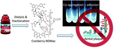
Neto CC, Penndorf KA, Feldman M, Meron-Sudai S, Zakay-Rones Z, Steinberg D, Fridman M, Kashman Y, Ginsburg I, Ofek I, et al. Characterization of non-dialyzable constituents from cranberry juice that inhibit adhesion, co-aggregation and biofilm formation by oral bacteria. Food & Function [Internet]. 2017;8 (5) :1955-1965. Publisher's VersionAbstract


An extract prepared from cranberry juice by dialysis known as nondialyzable material (NDM) has been shown previously to possess anti-adhesion activity toward microbial species including oral bacteria, uropathogenic Escherichia coli and Helicobacter pylori. Bioassay-guided fractionation of cranberry NDM was therefore undertaken to identify the anti-adhesive constituents. An aqueous acetone-soluble fraction (NDMac) obtained from Sephadex LH-20 inhibited adhesion-linked activities by oral bacteria, including co-aggregation of oral bacteria Fusobacterium nucleatum with Streptococcus sanguinis or Porphyromonas gingivalis, and biofilm formation by Streptococcus mutans. Analysis of NDMac and subsequent subfractions by MALDI-TOF MS and 1H NMR revealed the presence of A-type proanthocyanidin oligomers (PACs) of 3–6 degrees of polymerization composed of (epi)catechin units, with some (epi)gallocatechin and anthocyanin units also present, as well as quercetin derivatives. Subfractions containing putative xyloglucans in addition to the mixed polyphenols also inhibit biofilm formation by S. mutans (MIC = 125–250 μg mL−1). These studies suggest that the anti-adhesion activities of cranberry NDM on oral bacteria may arise from a combination of mixed polyphenol and non-polyphenol constituents.

 Characterization of non-dialyzable constituents from cranberry juice that inhibit adhesion, co-aggregation and biofilm formation by oral bacteria


food_function.pdf

Pages